Kamagra enthält Sildenafilcitrat als pharmakologisch aktiven Bestandteil. Dieser hemmt selektiv die Phosphodiesterase-5 und erhöht dadurch die Konzentration von cGMP im Corpus cavernosum. Der Effekt ist zeitlich begrenzt, da die Halbwertszeit von Sildenafil etwa vier Stunden beträgt. In der galenischen Form als Mundgel erfolgt die Resorption besonders rasch, was zu einem schnelleren Wirkeintritt führt. Der Abbau erfolgt überwiegend hepatisch über CYP3A4, wobei ein aktiver Metabolit entsteht, der zur Gesamtwirkung beiträgt. Typische Nebenwirkungen ergeben sich aus der Vasodilatation, darunter leichte Kopfschmerzen und nasale Kongestion. In klinischen Beschreibungen wird kamagra oral jelly im Zusammenhang mit der schnelleren Absorption erwähnt.
Genorise.cn
Bone morphogenetic protein 1 (BMP1), is a which in humans is encoded by the BMP1
(1, 2). There are seven isoforms of the protein created by . BMP1 belongs to thefamily of (BMPs). It induces bone and cartilagedevelopment. Unlike other BMPs, it does not belong to the superfamily. It was initially discoveredto work like other BMPs by inducing bone and cartilage development. It however, is a that cleaves the of and . It has an -like domain. It has beenshown to cleave and is localized in the basal epithelial layer of skin. The BMP1 locusencodes a protein that is capable of inducing formation of cartilage in vivo. Although other bonemorphogenetic proteins are members of the TGF-beta superfamily, BMP1 encodes a protein that is notclosely related to other known growth factors. BMP1 protein and procollagen C proteinase (PCP), asecreted metalloprotease requiring calcium and needed for cartilage and bone formation, are identical. PCP or BMP1 protein cleaves the C-terminal propeptides of procollagen I, II, and III and its activity isincreased by the procollagen C-endopeptidase enhancer protein. The BMP1 gene is expressed asalternatively spliced variants that share an N-terminal protease domain but differ in their C-terminalregion.
Tabas JA, Zasloff M, Wasmuth JJ, Emanuel BS, Altherr MR, McPherson JD, Wozney JM, Kaplan FS (February 1991). "Bone morphogenetic protein: chromosomal localization of human genes for BMP1, BMP2A, and BMP3". Genomics 9 (2): 283–9
2. Mac Sweeney A, Gil-Parrado S, Vinzenz D, Bernardi A, Hein A, Bodendorf U, Erbel P, Logel C, Gerhartz
B (December 2008). "Structural basis for the substrate specificity of bone morphogenetic protein 1/tolloid- like metalloproteases". J. Mol. Biol. 384 (1): 228–39.
This is a shorter ELISA assay that reduces time to 50% compared to the conventional method,
and the entire assay only takes 3 hours. This assay employs the quantitative sandwich enzymeimmunoassay technique and uses biotin-streptavidin chemistry to improve the performance andthe sensitivity of the assays. An antibody specific for human BMP1 has been pre-coated onto amicroplate. Standards and samples are pipetted into the wells and any BMP1 present is bound bythe immobilized antibody. After washing away any unbound substances, a detection antibodyspecific for human BMP1 is added to the wells. Following wash to remove any unboundantibody reagent, a detection reagent is added. After intensive wash a substrate solution is addedto the wells and color develops in proportion to the amount of BMP1 bound in the initial step. The color development is stopped and the intensity of the color is measured.
This package insert must be read in its entirety before using this product.
Copyright 2009-2013 GENORISE SCIENTIFIC, INC. Cat. 106189 www.genorise.com Page 1
Bring all reagents to room temperature before use. y Store at 4°C upon received. d (60 µL) – The Detection Antibody should be stored at -20°C to -
70°C in a manual defrost freezer for up to 6 months, if not used immediately. Centrifuge for 1 min at6000 x g to bring down the material prior to open the vial. The vial contains sufficient DetectionAntibody for a 96-well plate. If the volume is less than 60 µL, add 1 x Reagent Diluent to a final volumeof 60 µL and vortex briefly. Take 60 µL of detection antibody to 10 mL Reagent Diluent if the entire 96-well plate is used. If the partial antibody is used store the rest at -20°C until use. r (3 vials) – Human BMP1 Standard has a total of 3 vials. Each vial contains 75
µL of the standard sufficient for a 96-well plate. The undiluted standard can be stored at -20°C for up to 2months if not used immediately. Centrifuge for 1 min at 6000 x g to bring down the material prior to openthe tube. If the volume is less than 75 µL, add Standard Diluent to a final volume of 12 µL and vortexbriefly. Add 425 µL of Stamdard Diluent to one Standard vial containing 75 µL of the standard to makethe high standard concentration of 2600 pg /ml. Vortex briefly and allow it to sit for a minimum of 15 minprior to use. A seven point standard curve is generated using 2-fold serial dilutions in Standard Diluent,vortex briefly for each of dilution step. Store the rest of the standard at -20°C. n (50 µL) – Centrifuge for 1 min at 6000 x g to bring down the material prior to open the
vial. The vial contains sufficient Detection Antibody for a 96-well plate. If the volume is less than 50 µL,add 1 x Reagent Diluent to a final volume of 50 µL and vortex briefly. Make 1:200 dilutions in ReagentDiluent. If the entire 96-well plate is used, add 50 µL of Detection Agent to 10 mL Reagent Diluent priorto the assay. The rest of undiluted Detection Reagent can be stored at 4°C for up to 6 months. DO NOTFREEZE. B , pH 7.3, 30 mL- Dilute to 1 x PBS with deionized distilled water and mix well prior to use. e , 25 mL- Dilute to 1 x Wash Buffer with 1 x PBS prior to use. n , 3 mL – Prior to use dilute to 1 x Reagent Diluent with 1 x PBS and mix well. n , 15 mL – Prior to use dilute to 1 x Reagent Diluent with 1 x PBS and mix well.
Copyright 2009-2013 GENORISE SCIENTIFIC, INC. Cat. 106189 www.genorise.com Page 2
1. Vortex briefly the samples and the standards prior to the assay. Add 100 µL of sample (such as
plasma or serum) or standard to each well, cover the 96-well plate and incubate 1 hour at roomtemperature.
2. Aspirate each well and wash with 1 x Wash Buffer, repeating the process two times for a total of
three washes. Wash by filling each well with 1 x Wash Buffer (300 µL) using a multi-channelpipette, manifold dispenser or auto-washer. Complete removal of liquid at each step is essentialfor good performance. After the last wash, remove any remaining Wash Buffer by aspirating orby inverting the plate and blotting it against clean paper towels.
3. Add 100 µL of the working dilution of the Detection Antibody to each well. Cover the plate and
incubate 1 hour at room temperature.
4. Repeat the aspiration/wash as in step 2. 5. Add 100 µL of the working dilution of Detection Agent to each well. Cover the plate and
incubate for 20 minutes at room temperature. Avoid placing the plate in direct light.
6. Repeat the aspiration/wash as in step 2. 7. Add 100 µL of Substrate Solution to each well. Incubate for 10-20 minutes at room temperature.
Avoid placing the plate in direct light.
8. Add 50 µL of Stop Solution to each well. Gently tap the plate to ensure thorough mixing. 9. Determine the optical density of each well immediately, using a microplate reader set to 450 nm.
If wavelength correction is available, set to 540 nm or 570 nm. If wavelength correction is notavailable, subtract readings at 540 nm or 570 nm from the readings at 450 nm. This subtractionwill correct for optical imperfections in the plate. Readings made directly at 450 nm withoutcorrection may be higher and less accurate.
1. If BMP1 exceeds the upper limit of the detection, the sample needs to be diluted with 1 x Wash
Buffer. The dilution factor must be used for calculation of the concentration.
2. Detection Agent contains enzyme, DO NOT mass up with Detection Antibody. 3. The Stop Solution is an acid solution, handle with caution. 4. A standard curve should be generated for each set of samples assayed. 5. This kit should not be used beyond the expiration date on the label. 6. A thorough and consistent wash technique is essential for proper assay performance. Wash Buffer
should be dispensed forcefully and removed completely from the wells by aspiration or decanting. Remove any remaining Wash Buffer by aspiration or by inverting the plate and blotting it againstclean paper towels.
7. Use a fresh reagent reservoir and pipette tips for each step. 8. It is recommended that all standards and samples be assayed in duplicate. 9. Avoid microbial contamination of reagents and buffers. This may interfere with the sensitivity of
Copyright 2009-2013 GENORISE SCIENTIFIC, INC. Cat. 106189 www.genorise.com Page 3
Average the duplicate readings for each standard, control, and sample and subtract the
average zero (blank) standard optical density.
Create a standard curve by reducing the data using computer software capable of generating a
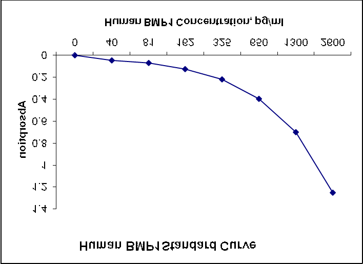
four parameter logistic (4-PL) curve-fit. As an alternative, construct a standard curve by plottingthe mean absorbance for each standard on the y-axis against the concentration on the x-axis anddraw a best fit curve through the points on the graph. The data may be linearized by plotting thelog of the BMP1 concentrations versus the log of the O.D. and the best fit line can be determinedby regression analysis. This procedure will produce an adequate but less precise fit of the data. Ifsamples have been diluted, the concentration read from the standard curve must be multiplied bythe dilution factor.
The graph below represents typical data generated when using this human BMP1 ELISA Kit.
The standard curve was calculated using a computer generated 4-PL curve-fit. For this case, aBio-Rad iMarkTM Microplate Reader and a Microplate Manager 6 Software were used togenerate this curve. The correlation coefficient (r2) is 0.999-1.000.
Copyright 2009-2013 GENORISE SCIENTIFIC, INC. Cat. 106189 www.genorise.com Page 4
The following recombinant human proteins prepared at 10 ng/ml were tested and exhibited no
cross-reactivity or interference. BMP2, BMP4, HGF, IL-1β, IL-2, IL-4, IL-5, IL-6, IL-8, IL-10, IL-12, IL-13, IL-15, IFNγ,MMP-2, TGFβ1, TGFβ2, TGFβ3, TLR1, TLR2, TLR3, TNF-α, VEGF.
This kit is calibrated against a highly purified CHO cell-expressed recombinant human BMP1.
20 x Sample Diluent, GERC-10305820 x PBS, Cat. 103004-2020 x ELISA Wash Buffer, Cat. 10302810 x ELISA Reagent Diluent, Cat. GERC-103055Universal Blocking Buffer, Cat.1030052 x Recombinant Protein Stabilizer, Cat. GERC-03014-25 x Recombinant Protein Stabilizer, Cat. GERC-103014-5ELISA G-Blue Substrate Solution, Cat. 103021Human BMP1 StandardHuman BMP1 detection antibody
Copyright 2009-2013 GENORISE SCIENTIFIC, INC. Cat. 106189 www.genorise.com Page 5
An a m n e s e b o g e n Bitte helfen Sie uns, damit wir Ihnen bei der Behandlung die bestm€gliche medizinische Betreuung zukommen lassen k€nnen. Um gesundheitliche Risiken zu vermeiden, ist es wichtig, dass Sie alle folgenden Fragen gewissenhaft beantworten. Mit Ihren Antworten gehen wir vertraulich um. Sie unterliegen der •rztlichen Schweigepflicht und dienen ausschlie‚lich de
ADULT PERITONEAL DIALYSIS PATIENTS STANDING ORDERS 1. ALL PATIENTS TREATED WITH PERITONEAL DIALYSIS 1. PO water-soluble vitamin replacement daily. 2. Colace or equivalent, 100 mg., p.o. BID, unless diarrhea. 3. Gentamicin 0.1% cream to PD catheter exit site daily. Influenza vaccine should be administered to all patients except those with egg allergy, those for whom the patient’s physician
 Bone morphogenetic protein 1 (BMP1), is a which in humans is encoded by the BMP1
(1, 2). There are seven isoforms of the protein created by . BMP1 belongs to thefamily of (BMPs). It induces bone and cartilagedevelopment. Unlike other BMPs, it does not belong to the superfamily. It was initially discoveredto work like other BMPs by inducing bone and cartilage development. It however, is a that cleaves the of and . It has an -like domain. It has beenshown to cleave and is localized in the basal epithelial layer of skin. The BMP1 locusencodes a protein that is capable of inducing formation of cartilage in vivo. Although other bonemorphogenetic proteins are members of the TGF-beta superfamily, BMP1 encodes a protein that is notclosely related to other known growth factors. BMP1 protein and procollagen C proteinase (PCP), asecreted metalloprotease requiring calcium and needed for cartilage and bone formation, are identical.
Bone morphogenetic protein 1 (BMP1), is a which in humans is encoded by the BMP1
(1, 2). There are seven isoforms of the protein created by . BMP1 belongs to thefamily of (BMPs). It induces bone and cartilagedevelopment. Unlike other BMPs, it does not belong to the superfamily. It was initially discoveredto work like other BMPs by inducing bone and cartilage development. It however, is a that cleaves the of and . It has an -like domain. It has beenshown to cleave and is localized in the basal epithelial layer of skin. The BMP1 locusencodes a protein that is capable of inducing formation of cartilage in vivo. Although other bonemorphogenetic proteins are members of the TGF-beta superfamily, BMP1 encodes a protein that is notclosely related to other known growth factors. BMP1 protein and procollagen C proteinase (PCP), asecreted metalloprotease requiring calcium and needed for cartilage and bone formation, are identical. Bring all reagents to room temperature before use.
 1. Vortex briefly the samples and the standards prior to the assay. Add 100 µL of sample (such as
plasma or serum) or standard to each well, cover the 96-well plate and incubate 1 hour at roomtemperature.
1. Vortex briefly the samples and the standards prior to the assay. Add 100 µL of sample (such as
plasma or serum) or standard to each well, cover the 96-well plate and incubate 1 hour at roomtemperature.
 Average the duplicate readings for each standard, control, and sample and subtract the
average zero (blank) standard optical density.
Average the duplicate readings for each standard, control, and sample and subtract the
average zero (blank) standard optical density. The following recombinant human proteins prepared at 10 ng/ml were tested and exhibited no
cross-reactivity or interference.
The following recombinant human proteins prepared at 10 ng/ml were tested and exhibited no
cross-reactivity or interference.