Intranet.karolinskaeducation.ki.se

Effect of joint injections in children with juvenile idiopathic arthritis:evaluation by 3D-gait analysis E Brostro¨m, S Hagelberg and Y Haglund-A Department of Woman and Child Health, Karolinska Institute, Astrid Lindgren Children’s Hospital, Stockholm, Sweden ˚ kerlind Y. Effect of joint injections in children with juvenile idiopathic arthritis: evaluation by 3D-gait analysis. Acta Pædiatr 2004; 93: 906–910. Stockholm.
ISSN 0803-5253 Aim: To investigate how gait patterns change after intra-articular corticosteroid injections (ICIs) inthe lower extremities. Methods: Eighteen children, aged 5–16 y, with juvenile idiopathic arthritis(JIA) and lower extremity involvement participated in the study. Sixty-four joints in the lowerextremities were treated with ICIs. The Visual Analogue Scale (VAS, 0–100 mm) was used toassess pain. A 3D motion analysis system and two force plates were used to measure gaitparameters, kinematics and kinetics. The first gait analysis was performed before treatment withICIs, and a second one was done 8–17 d after treatment. Results: The participants’ average ratingof pain decreased from 26 mm on the VAS before ICI to 11 mm (p = 0.001) after treatment. Theself-chosen walking velocity was significantly faster (p = 0.02) after treatment. The range of kneeand ankle joint angles during gait increased significantly (p = 0.03 and 0.04) after treatment. Atloading response, the hip extension moment increased (p = 0.01) as did knee flexion moment, andplantar flexion moment at pre-swing increased significantly (p = 0.02 and 0.002) after treatment.
The ankle also generated more power (p = 0.005) after treatment.
Conclusion: The study shows positive effects of treatment with ICIs in the lower extremities—especially regarding pain, walking velocity and joint moments. The data indicate that ICItreatment influences the gait pattern also in joints that have not been injected.
Key words: Gait, juvenile rheumatoid arthritis, kinematics, kinetics, locomotion
Eva Brostro¨m, Department of Woman and Child Health, Astrid Lindgren Children’s Hospital,Q2:07, SE-171 76 Stockholm, Sweden (Tel. ‡46 8 5177 7636, fax. ‡46 8 5177 7351, e-mail.
Eva.brostrom@kbh.ki.se)
The lower extremities are often affected in juvenile and leg length (9, 12–14). Various studies have shown idiopathic arthritis (JIA). This can lead to functional that children with JIA experience pain and stiffness disabilities, with deviations from the normal gait together with reduced physical capability and altered pattern, including restricted joint motion, velocity, limping and reduction in step length. Long duration of Three dimensional (3D) gait analyses, including active disease is also associated with a reduction in kinematics and kinetics, provide more information height (1–4), discrepancy in leg length, and shortening about gait changes, such as joint angles and moments, of muscle and tendon length that causes joint flexion which are impossible to quantify in a standard clinical contractures (5–7). Early use of intra-articular cortico- setting. The kinematics shows the joint movement, and steroid injections (ICIs) in the joint rapidly resolves the kinetics describes the forces involved in movement synovitis, protects against joint deformity and alleviates (e.g. ground reaction forces, joint moments, and joint pain, thus creating a good opportunity for physiotherapy powers). By examining kinetics, the mechanisms of gait and rehabilitation (8). Furthermore, by using ICIs, leg- deviation can be described. Gait analysis has the length discrepancies may be avoided, which, in turn, potential to measure pathological changes in ambula- reduces the need for systemic treatment with steroids tion (15, 16) and can provide important information about children’s gait patterns, thus helping physicians to Walking is a task that we seek to understand better make the correct decisions regarding treatments that because it is one of the most fundamental physical include, for example, ICI and physical therapy (12, 17, functions of the human being. In earlier studies by 18). Early use of gait analysis can be instrumental in others and by ourselves (10, 11) it was identified that discovering developments of potentially destructive gait children with JIA generally walked more slowly than their healthy peers, that walking velocity varies with The aim of this study was to determine the effects of age, and, more importantly, with factors such as height ICI treatment in the lower limbs by using 3D gait  2004 Taylor & Francis. ISSN 0803-5253 Joint injections in children with JIA Table 1. Demographic data showing mean (SD) for children with JIA 
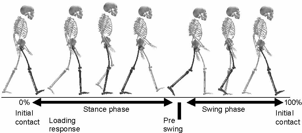
England). Thirty-four reflective markers (25 mm) were attached bilaterally on the subject’s skin at the head,shoulders, arms, pelvis, legs and feet according to the biomechanical gait model (Plugin Gait, Vicon Motion Systems). A total of three completed walking trials were performed for the kinematics and time-distance par- ameters. Two force plates (Kistler, Basel, Switzerland) along the walkway were used to measure ground reaction forces to produce joint kinetic information using inverse dynamics. One to three completed walking trials were performed to measure kinetics. A walking trial was considered completed if the subject’s right or left foot made a clean contact on the force plate.
The children were advised not to walk for 24 h and to avoid physical exercise for one week after ICItreatment. The second gait analysis was performed 8–17 d after treatment with ICI. To imitate the conditions analysis to describe joint angles and kinetics during at the first gait analysis, the same procedure was locomotion in the hip, knee and ankle.
followed and the same examiner (EB) performed theclinical measurements and marker placements.
Kinematics, kinetics, and time-distance parameters Eighteen children aged between 5 and 16 y with JIA were derived using the Vicon PlugIn Gait model. The participated in the study. All subjects had been lower body was modelled as 7 segments (pelvis, 2 diagnosed according to the International League thighs, 2 shanks, 2 feet). Ground reaction forces, Against Rheumatism (ILAR) classification system moments and power were normalized to body weight.
(20), and suffered from lower extremity involvement Stride parameters were normalized to height. An and polyarthritis. All patients were recruited from the ensemble, or point-to-point average gait cycle was clinic of Paediatric Rheumatology, Astrid Lindgren generated, from which gait patterns of the hip, knee and Children’s Hospital, Karolinska Hospital in Stockholm.
foot joint angles and joint moments were derived both Two patients were excluded because of medical before and after treatment with ICI. Joint power was conditions that prevented them from returning for the defined as the scalar product of the moment and the joint second gait analysis. The participants were independent angular velocity, and an entire gait cycle was defined walkers and had been injected with methylprednisolone from first contact to foot contact of the same limb acetate (Depo-medrol2) in 1 to 10 joints in the lower extremities. Our policy is to inject any joint that is The following peak values from the subjects’ active (with synovitis). During the study, 64 active ensemble gait cycle were analysed: hip flexion and joints were treated uni- or bilaterally with ICI (Table 1).
extension; maximum hip flexion, extension and abduc- The subjects’ characteristics are presented in Table 1.
tion moments; maximum knee flexion and extension; The database from our gait laboratory with 21 healthy maximum knee flexion, extension and varus moments; children between 5 and 14 y of age functioned as maximum ankle plantar flexion and dorsiflexion angles; controls when interpreting the data. The Research maximum plantar flexion and dorsiflexion moments; Ethics Committee of the Karolinska Hospital approved maximum power generation and absorption at the ankle.
the study. Verbal and written information was given tothe children and their parents.
ProcedureAll the children were asked to rate their level of pain ona visual analogue scale (VAS, 0–100 mm, using amoveable bar) prior to, during, and after the procedurein order to record increased pain. VAS has proven to bea reliable tool for measuring the intensity of pain inchildhood (21). The subjects walked barefoot on a 7.5-m-long walkway at a self-chosen velocity. Movementrecordings were done with a 6-camera 3D motion Fig. 1. Normal gait cycle defined from initial heel contact to heel analysis system (Vicon, Motion System, Oxford, Table 2. Mean values (SD) for gait parameters pre- and post- and velocity before and after treatment. All time- treatment with intra-articular corticosteroid injections in children distance parameters are presented in Table 2.
Hip joint angles. The hip range of motion (maximum hip flexion to maximum extension) did not show any significant difference after treatment. The average was 38° before and after ICI (range 14°–55°).
JIA: juvenile idiopathic arthritis; ICI: intra-articular corticosteroid Hip moments. The hip extension moment, which takes place during the loading response (the first 5–10% of thegait cycle), increased significantly (p = 0.01) (Table 3)after treatment with ICI, even if the subjects were not treated in the hip joint. There were no significant A two-way analysis of variance (ANOVA), repeated differences between left and right hip joint moments measures design, was used to assess the kinematics and and there was no significant difference in peak hip kinetic data from the 3D-gait analysis. The factors were flexion moment after treatment (Table 3). Two children side (left and right) and treatment (before and after ICI).
with JIA were injected bilaterally in the hip joint and For time and stride variables, non-parametric statistics two were injected only in the right hip (Table 3).
(Wilcoxon’s matched pairs test) were used to test forsignificance. Friedman ANOVA by rank test was used Knee joint angles. The knee range of motion (maximum to identify pain (VAS) before and after ICI treatment.
knee extension to maximum flexion) during gait The level of significance was set to p  0.05. All increased significantly (p = 0.03) from 50° (range 33°– statistical analyses were performed using Statistica 6.0 63°) to 54° (range 39°–63°) after treatment.
Knee moments. Of the entire group of children with JIAthat were injected in the lower extremities, none of the subjects showed a significant difference in peak knee extension moments at loading response after treatment The participants rated their pain before the ICI at 26 mm with ICI (Table 3). There was a significant increase (median value, range 0–69 mm) on the VAS. After (p = 0.02) in peak knee flexion moment at pre-swing (50% of the gait cycle) after treatment with ICI. There (p = 0.001) reduced to 11 mm (range 0–32 mm).
were no significant differences between left and rightknee joints. No significant differences were shown inthe frontal moment at initial contact and at pre-swing at horizontal level after treatment with ICI in the knee The participants showed a significantly faster self- chosen walking velocity after treatment with ICI Seven subjects were injected with ICI in the knee (p = 0.02). There was no correlation between pain rating joint, five bilaterally and two unilaterally (Table 3).
Table 3. Mean joint moments (units are Nm/kg) in the sagittal plane. These refer to mean values for the whole JIA population (SD) and theinjected joints before and after ICI treatment and non-injected joints. Data were obtained from both the left and right gait cycles.
Joint injections in children with JIA Ankle joint angles. The ankle joint angle range (maxi- two other studies (27, 28) it has been shown that mum dorsiflexion to maximum plantar flexion) during children with JIA have a moderate to large impairment gait increased significantly (p = 0.04) from 19° to 22° in their cardio-respiratory fitness compared with healthy children. Knowing this, it is important to create exerciseopportunities for children with JIA so that they can Ankle moments. The ankle plantar flexion moment at participate in physical activities and sports.
pre-swing showed a significant difference (p = 0.002) Children with JIA have been reported to use lower while dorsiflexor moment at loading response did not self-chosen walking velocity, with reduced vertical change after treatment with ICI (Table 3).
ground reaction forces at heel strike and push off, Thirteen children were injected in the right foot and compared with healthy, age-matched controls (9). Other 11 were injected in the left foot (Table 3); 8 children studies have pointed out the importance of normalizing were injected unilaterally and 8 were injected bilaterally walking velocity to height to allow comparisons between children of the same age with and withoutJIA (10, 11, 26). In their study, Fairburn et al. (19) found Power. Ankle power increased significantly (p = 0.005) that children with JIA showed abnormal gait patterns at 50% of the gait cycle (stance phase) after treatment even though the joint activities were low.
The 3D-gait analysis data showed increased range between maximum flexion and extension angles duringlocomotion in the knee and ankle. This is important because it indicates that the children with JIA have theopportunity to change their ingrained gait patterns.
This study has shown the positive effects of treatment Children and adults with arthritis often suffer from foot with ICI in the lower extremities—especially on pain, problems, categorized as pronated foot, synovitis, and walking velocity and joint moments. Furthermore, and have a diminished range of motion in the ankle and toe very importantly, the data indicates that ICI treatment valgus (29). This indicates that foot problems are influences the gait pattern even in joints not being common in the JIA population, underscoring the need treated. The aim of this study was to evaluate gait after for thorough evaluation and management of physical treatment, using the patients as their own controls and not to compare them with untreated children with JIA or The hip extension joint moment at loading response, knee flexion joint moment at pre-swing and ankle To our knowledge, this is the first study that has been plantar flexion joint moments at stance showed in- undertaken to evaluate gait pattern after treatment with creased values after treatment with ICI. However, we ICI. Three-dimensional gait analysis is a well-estab- found that all joint moments in children with JIA lished method enabling highly objective and reliable showed decreased values in comparison with those of evaluation of gait in both healthy and diseased popula- healthy children in our database. This study indicates tions (12, 18, 22). It is also important to bear in mind that the plantar flexor moment increased after treatment that the pharmaceutical methyl-prednisolone acetate with ICI, which is important in order to manage toe-off.
(Depo-medrol2) we used does not have as lasting an The main muscle groups that are active at the ankle effect as triamcinolone hexacetonide (Lederspan2) throughout stance are the plantar flexors (triceps surae).
(23), which was not available on the market at the time We know from our earlier studies (30) that children with of this study. Our clinical experience is that ICIs in JIA are weaker in the plantar flexors compared with general have a good and prompt effect on the treated joints. The gait pattern of children with JIA is com- To evaluate the muscle action during a movement, plicated by compensatory gait alternations, resulting we also need to calculate the power, which tells us how from joint pain, stiffness, and in some cases joint the muscle group is contracting (isometrically, concen- deformity. It has been shown that the kinematics of the trically or eccentrically). It does not, however, tell us in hip, knee and ankle joints is clearly related to walking detail how the muscle contracts. When we assessed the velocity (24), and that both the profile and magnitude of power (W/kg) we found that the hip and knee values the external ground reaction forces are velocity depen- were almost normal (compared with healthy children in dent (25, 26). A higher walking velocity generally our database), while the ankle powers were decreased results in elevated vertical and anterior–posterior (2.04 watt vs 2.95 watt) in children with JIA.
ground reaction forces, which consequently require In conclusion, this group of children with JIA showed greater lower-limb muscle strength. The participant’s significant positive changes in their gait patterns after self-chosen walking velocity in this study increased treatment with ICI in the lower extremities measured by significantly after treatment, which is important for 3D-gait analysis. These results point out how important daily activities. There was no relationship between pain it is for physicians to use ICI treatment, which is rating and velocity in this study, perhaps because the preferable to systemic steroids. Furthermore, this group group was too small to show statistical correlation. In of children was able to absorb and generate more muscle power after ICI. All this information is of spatial parameters of gait in children. II: Pathological gait. Dev interest in the interpretation of walking. This is clinically important because the disease often affects 14. Wheelwright EF, Minns RA, Law HT, Elton RA. Temporal and spatial parameters of gait in children. I: Normal control data. Dev daily activities such as walking and running, and sometimes has a negative impact on physical ability.
15. Ramakrishnan HK, Kadaba MP. On the estimation of joint Since the moments and powers cannot be measured kinematics during gait. J Biomech 1991; 24: 969–77 without 3D-gait analysis, this advanced technique could 16. Kadaba MP, Ramakrishnan HK, Wootten ME. Measurement of lower extremity kinematics during level walking. J Orthop Res be used throughout the disease periods accurately to evaluate treatment, gait patterns and progress.
17. Perry J. Gait analysis: technology and the clinician. J Rehabil Res Acknowledgements.—The study was funded by grants from The 18. Sutherland DH, Olshen R, Cooper L, Woo SL. The development Swedish Rheumatism Association, the Vardal Foundation for Health of mature gait. J Bone Joint Surg Am 1980; 62: 336–53 Care Science and Allergy Research, the Foundation Frimurare 19. Fairburn PS, Panagamuwa B, Falkonakis A, Osborne S, Palmer Barnhuset in Stockholm, the Sunnerdahl and Norbacka Foundation, R, Johnson B, et al. The use of multidisciplinary assessment and the Society for Child Care and the Solstickan Foundation.
scientific measurement in advanced juvenile idiopathic arthritiscan categorise gait deviations to guide treatment. Arch Dis Child2002; 87: 160–5 20. Petty RE, Southwood TR, Baum J, Bhettay E, Glass DN, Manners P, et al. Revision of the proposed classification criteriafor juvenile idiopathic arthritis: Durban, 1997. J Rheumatol 1. Bacon MC, White PH, Raiten DJ, Craft N, Margolis S, Levander OA, et al. Nutritional status and growth in juvenile rheumatoid 21. McGrath PJ. Annotation: aspects of pain in children and arthritis. Semin Arthritis Rheum 1990; 20: 97–106 adolescents. J Child Psychol Psychiatry 1995; 36: 717–30 2. Cassidy JT, Petty RE. Textbook of pediatric rheumatology. 2nd 22. Kadaba MP, Ramakrishnan HK, Wootten ME, Gainey J, Gorton ed. New York: Churchill Livingstone Inc.; 2002 G, Cochran GV. Repeatability of kinematic, kinetic, and electro- 3. Hagelberg S, Magnusson B, Andersson U. Improved growth in myographic data in normal adult gait. J Orthop Res 1989; 7: 849– children with juvenile idiopathic arthritis (JIA) treated with etanercept. Ann Rheum Dis 2001; 60 Suppl II 23. Martini G, Gobber D, Agosto C, Vianello A, Zulian F. Com- 4. Schneider R, Passo MH. Juvenile rheumatoid arthritis. Rheum parison between intraarticular triamcinolone hexacetonide and acetonide in oligoarticular JIA. Ann Rheum Dis 2001; 60 Suppl 5. Fan JS, Wessel J, Ellsworth J. The relationship between strength and function in females with juvenile rheumatoid arthritis. J 24. Nilsson J, Thorstensson A. Adaptability in frequency and ampli- tude of leg movements during human locomotion at different 6. Lindehammar H, Backman E. Muscle function in juvenile speeds. Acta Physiol Scand 1987; 129: 107–14 chronic arthritis. J Rheumatol 1995; 22: 1159–65 25. Nilsson J, Thorstensson A. Ground reaction forces at different 7. Vostrejs M, Hollister JR. Muscle atrophy and leg length speeds of human walking and running. Acta Physiol Scand 1989; discrepancies in pauciarticular juvenile rheumatoid arthritis.
26. van der Linden ML, Kerr AM, Hazlewood ME, Hillman SJ, 8. Henderson CJ, Lovell DJ, Specker BL, Campaigne BN. Physical Robb JE. Kinematic and kinetic gait characteristics of normal activity in children with juvenile rheumatoid arthritis: quantifica- children walking at a range of clinically relevant speeds. J Pediatr tion and evaluation. Arthritis Care Res 1995; 8: 114–9 9. Sherry DD, Stein LD, Reed AM, Schanberg LE, Kredich DW.
27. Takken T, van der Net PJM, Helders PJM. Do juvenile idiopathic Prevention of leg length discrepancy in young children with arthritis patients benefit from an exercise program? A pilot study.
pauciarticular juvenile rheumatoid arthritis by treatment with intraarticular steroids. Arthritis Rheum 1999; 42: 2330–4 28. Takken T, van der Net PJM, Helders PJM. Aerobic exercise 10. Brostrom E, Haglund-Akerlind Y, Hagelberg S, Cresswell AG.
testing in juvenile rheumatoid arthritis (JRA) patients. Clin Gait in children with juvenile chronic arthritis. Timing and force parameters. Scand J Rheumatol 2002; 31: 317–23 29. Spraul G, Koenning G. A descriptive study of foot problems in 11. Frigo C, Bardare M, Corona F, Casnaghi D, Cimaz R, Naj Fovino children with juvenile rheumatoid arthritis (JRA). Arthritis Care P, et al. Gait alteration in patients with juvenile idiopathic arthritis: a computerized analysis. J Orthop Rheumatol 1996; 9: 30. Hedengren E, Knutson LM, Haglund-Akerlind Y, Hagelberg S.
Lower extremity isometric joint torque in children with juvenile 12. Ounpuu S, Gage JR, Davis RB. Three-dimensional lower chronic arthritis. Scand J Rheumatol 2001; 30: 69–76 extremity joint kinetics in normal pediatric gait. J Pediatr Orthop1991; 11: 341–9 Received July 11, 2003; revisions received Sept. 18, 2003; accepted 13. Wheelwright EF, Minns RA, Elton RA, Law HT. Temporal and

Source: http://intranet.karolinskaeducation.ki.se/files/978/Joint%20incections%20JIA%20EB.pdf

Entre el repique y el estruendo, la celebración del 5 de mayo en puebla, 1868-1930

Entre el repique y el estruendo, la celebración del 5 de Mayo en Puebla, 1868-1930 Rosalina Estrada Urroz - Enrique Cano Galindo Entre el repique y el estruendo, la celebración del 5 de Mayo en Puebla, 1868-1930 Date de mise en ligne : samedi 19 janvier 2013 Description : México, Imperio de Maximiliano, Fiesta, Conmemoración, 5 de Mayo 1862 Artelogie Entre el repique y

elasten.it

PRATO, 24/06/2012 Elasticized linen, the patent comes from Prato Elasten is the company which produced it. A few weeks ago the patent application. Now the market response is expected. Its code name is WO 2012/056436A2, actually it's nothing but a yarn. A yarn that might reveal very precious and be included in the list of inventions that have made Prato the textile capital of Europe. The pat

Copyright © 2010-2014 Online pdf catalog