Kamagra enthält Sildenafilcitrat als pharmakologisch aktiven Bestandteil. Dieser hemmt selektiv die Phosphodiesterase-5 und erhöht dadurch die Konzentration von cGMP im Corpus cavernosum. Der Effekt ist zeitlich begrenzt, da die Halbwertszeit von Sildenafil etwa vier Stunden beträgt. In der galenischen Form als Mundgel erfolgt die Resorption besonders rasch, was zu einem schnelleren Wirkeintritt führt. Der Abbau erfolgt überwiegend hepatisch über CYP3A4, wobei ein aktiver Metabolit entsteht, der zur Gesamtwirkung beiträgt. Typische Nebenwirkungen ergeben sich aus der Vasodilatation, darunter leichte Kopfschmerzen und nasale Kongestion. In klinischen Beschreibungen wird kamagra oral jelly im Zusammenhang mit der schnelleren Absorption erwähnt.
Correctiezolen.nl
David D. Dyck, Jr., DO,* and Lori A. Boyajian-O’Neill, DO, FAOASM*†
dial tubercle of the calcaneus. The plantar fascia extends from
(Clin J Sport Med 2004;14:305–309)
this tubercle to the metatarsal heads, forming the longitudinalarch that provides support for the foot. Excessive load or ten-sion on this aponeurosis can lead to the condition commonly
Plantarfasciitisaffectsanestimated2millionpersonswho referredtoasplantarfasciitis.Historically,plantarfasciitishas
present annually for medical treatment.1 Runners comprise
been considered an inflammatory process. However, in recal-
over 76% of athletes with this condition.2,3 Men and women
citrant cases, it appears that a degenerative process affecting
appear to be equally affected.4 Plantar fasciitis classically
collagen5 or degenerative fasciosis10 occurs without evidence
presents as medial heel pain that is sharp and most severe with
of inflammation. Lemont et al10 performed histologic exami-
the first step out of bed in the morning or after prolonged rest.
nation of surgical biopsies, revealing increased fibroblasts,
This condition is generally self-limiting, but complete resolu-
ground substance, and vascularity, not the expected inflamma-
tion may take 6 to 18 months or longer.5 Aggressive manage-
tory mediators.5,10 This improved understanding may impact
ment using combination therapies is most efficacious and af-
the use of anti-inflammatory therapies for chronic plantar
fords athletes the best opportunity to recover quickly and fully.
An understanding of risk factors and pathophysiology willhelp in identifying athletes predisposed to developing plantar
TREATMENT
fasciitis, as well as in formulating the most effective preven-
Treatment should be aggressive, should be initiated as
tive and therapeutic treatment plan.
early as possible, and should include a multifaceted approachthat addresses underlying anatomic and biomechanic condi-
RISK FACTORS
tions, training errors, and risk factors. Assessment of underly-
Plantar fasciitis can occur acutely, as with trauma, but
ing risk factors is the foundation for successful treatment and
more often presents as chronic plantar foot pain of insidious
prevention. Treatment options discussed are summarized in
onset associated with chronic overload. In approximately 85%
of patients, the etiology is undetermined.6,7 Risk factors for
Relative Rest
developing plantar fasciitis can be divided into anatomic, bio-
Relative rest must be a part of the treatment plan, allow-
mechanical, and environmental and are presented in Table 1.
ing athletes to continue to train in some capacity without ex-
Limited ankle dorsiflexion (Յ10°) has been reported as the
acerbating symptoms. Relative rest relieves microtrauma
most important independent risk factor.8 This limitation may
caused by repetitive overload. Wolgin et al11 reported 25% of
cause compensatory excessive pronation of the subtalar joint,
patients citing rest as the treatment that best relieved pain.
which increases the tensile load to the plantar aponeurosis.9
Cross-training should be considered to maintain aerobic fit-ness levels. Relieving the overuse component through rest is
PATHOPHYSIOLOGY
critical to the prevention of further injury.
Plantar fasciitis has been referred to as heel pain syn-drome and heel spur syndrome, because the pain is usually
Stretching
localized to the insertion of the plantar aponeurosis at the me-
Stretching is the single most effective method of treat-
ment. Of the 83% of patients who improved with stretching,29% indicated that stretching was most effective in relieving
Received for publication February 2004; accepted May 2004.
pain as compared with other modalities.11 Stretching should
From the *Department of Family Medicine, Kansas City University of Medi-
cine and Biosciences, Kansas City, MO; and †Primary Care Sports Medi-
include techniques targeted at the entire lower extremity, es-
cine, Kansas City University of Medicine and Biosciences, Kansas
pecially the plantar fascia and gastrocsoleus-Achilles com-
Reprints: David D. Dyck, Jr., DO, Kansas City University of Medicine and
The athlete can target the plantar fascia at home using a
Biosciences, 1750 Independence Avenue, Kansas City, MO 64106-1453
tennis ball, golf ball, or 15-oz can rolled under the arch to pro-
Copyright 2004 by Lippincott Williams & Wilkins
vide an adequate stretch. To accentuate further the stretch on
Clin J Sport Med • Volume 14, Number 5, September 2004
Clin J Sport Med • Volume 14, Number 5, September 2004
TABLE 1. Classification of Risk Factors for Plantar Fasciitis Anatomical Risk Factors Biomechanical Risk Factors Environmental Risk Factors
the fascia, the athlete can rest the affected foot on the contra-
use may decrease duration of symptoms, and they can be pur-
lateral or unaffected thigh, applying firm dorsiflexion of the
chased without a prescription. Powell et al13 reported that 88%
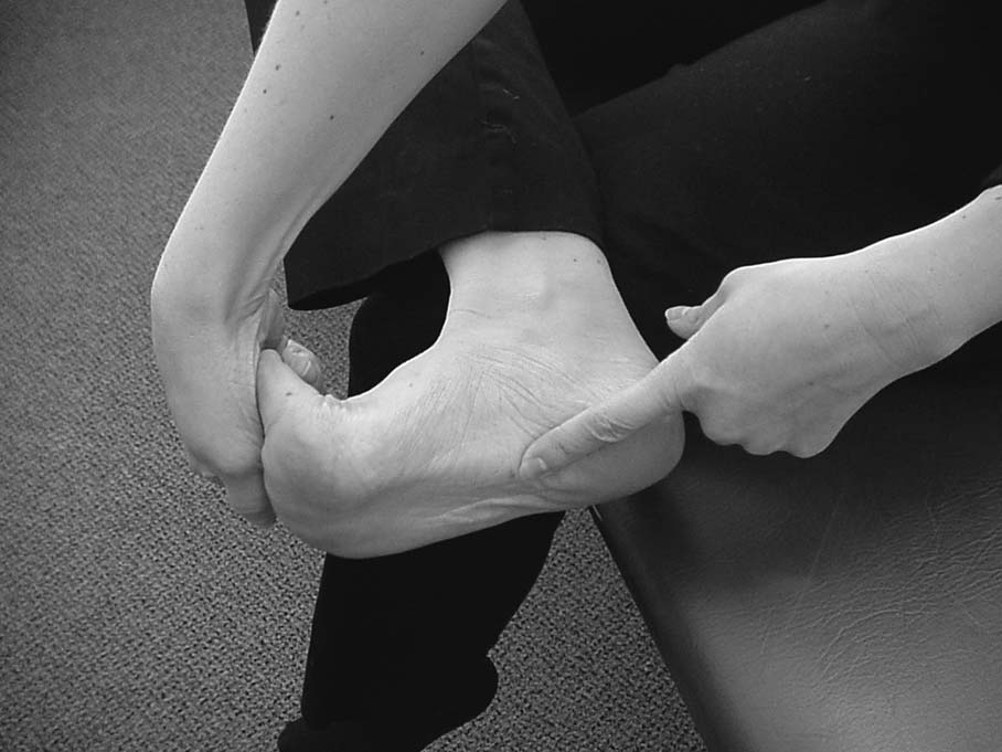
great toe, thus increasing the stretch of the plantar fascia (Fig.
of patients using night splints for 1 month experienced im-
1).12 Manual stretching techniques such as myofascial release
provement in their symptoms. They can be bulky and uncom-
and deep tissue massage can be used to stretch the fascia.
fortable but may return the athlete to peak performance more
Stretching the gastrocsoleus-Achilles complex is
quickly. Frequently, night splints are reserved for recalcitrant
achieved using wall stretches with a straight knee to isolate the
plantar fasciitis; we propose that they be considered at the on-
gastrocnemius muscle and with a bent knee to target the soleus
muscle. These stretches can also be performed using a curb orstair. Stretching techniques focusing on the plantar fascia has
Strengthening
been shown to accelerate recovery time and is more effective
Plantar flexor muscle strength deficits were cited by
than those directed exclusively at the gastrocsoleus-Achilles
Kibler et al14 as contributing to plantar fasciitis. Martin et al15
complex.12 Early prescriptions should be given for a home
reported that strengthening exercises provide the greatest de-
stretching program and possibly for physical therapy referral.
crease in pain in 34.9% of patients with plantar fasciitis.
Specific stretching techniques appear in Table 2.
Strengthening exercises for the intrinsic muscles of the foot aredesigned to improve longitudinal arch support and decrease
Night Splints
Dorsiflexion night splints (90°) relieve pain by provid-
Athletes can perform strengthening exercises every hour
ing continuous passive stretching during rest and sleep. Their
throughout the day by simple tapping of the toes with the footplanted. The desired technique is to raise the toes and pressthem each individually to the floor. Additionally, with a towelplaced on the floor, the athlete is instructed to keep the heelplanted and gather the towel by curling the toes. As strengthimproves, weight can be added to the towel to increase resis-tance.
Strengthening of the gastrocsoleus-Achilles complex is
accomplished using heel raises. The athlete begins with bothlegs at once and progresses to single leg repetitions. Asstrength improves, resistance can be increased using dumb-bells or free weights. We recommend that athletes performstrengthening exercises 3 times per day with 12 to 15 repeti-tions per set. Pain should be monitored with modifications infrequency and intensity to avoid exacerbation or return ofsymptoms. Anti-inflammatory Therapies
Recent histologic evidence identifying collagen degra-
FIGURE 1. Effective stretching of the plantar fascia accom-
dation, as opposed to inflammatory markers in surgical biopsy
plished by applying firm dorsiflexion of the great toe. Leftindex finger points at the medial aspect of the stretched fascia.
specimens, raises questions about the utility of anti-
2004 Lippincott Williams & WilkinsClin J Sport Med • Volume 14, Number 5, September 2004
TABLE 2. Treatment Options for Plantar Fasciitis Treatment Comments
Plantar fascia: 15-oz can rolled under arch, cross-friction
Decrease tension of gastrocsoleus-Achilles complex
massage, great toe extension, towel stretch
Gastrocsoleus: slant board, wall stretch, curb or stair stretch
Commercially available, compliance difficult
Improve structural integrity of longitudinal arch
Decrease inflammation, local pain control
Time-consuming, reserve for elite athletes or laborers
Decrease inflammation, local pain control
Use in later stages, risk of plantar fascia rupture, atrophy
Mild pes planusAdolescents experiencing rapid growthSymptoms less than 8 weeks
Correct anatomical and biomechanical factors
Change shoes every 300–500 milesCheck for correctable problems
High-energy: single treatment, local anesthesia needed
Low-energy: multiple treatment sessions, no anesthesianeeded, standardization still needed
Failed conservative therapy at least 6 months, often much
inflammatory therapy such as NSAIDs for chronic plantar
plantar fasciitis. Studies have shown a 70% success rate using
fasciitis.5,10 While NSAIDs are effective in some patients and
steroid injections when applied early in the disease process.7,18
were reported in 1 study to be the most effective treatment by
Injections can be performed using a plantar or medial approach
11% of subjects,11 the use of NSAIDs should be limited to a
with or without ultrasound guidance. Complications include
brief duration. Specifically, this may be 1 to 2 weeks at a time
rupture of the plantar fascia and fat pad atrophy. Although rup-
ture of the plantar fascia is uncommon, injection of corticoste-
Ice applied to the attachment of the aponeurosis follow-
roids has been suggested as a contributing factor.3,19,20 Most
ing activity can decrease pain and inflammation. At home, the
patients had resolution of symptoms associated with rupture
athlete can use ice massage to stretch the fascia. Iontophoresis
within 6 to 8 weeks.3,21 Still, the use of corticosteroid injec-
can be used to deliver corticosteroid such as dexamethasone to
tions in plantar fasciitis remains controversial.
the deep plantar aponeurosis. Gudeman et al reported improve-ment after 2 weeks but no long-term difference at 6 weeks.16,17
Arch Supports
Difficulties with this treatment are the time requirement and
The longitudinal arch is designed to distribute forces
expense, as optimal therapy requires administration 2 to 3
generated at heel strike. Anatomic, biomechanical, and envi-
times per week by a qualified professional. Recommendations
ronmental factors causing abnormal distribution of these
are to reserve iontophoresis for elite athletes and laborers pre-
forces can result in plantar fasciitis. Arch supports, through
taping or orthoses, can alter the transmission of forces and de-
Corticosteroid injections are controversial and are not a
crease stress. As a simple noninvasive treatment, they can be
first-line therapy due to the associated risks and possible com-
considered a first-line treatment of plantar fasciitis in combi-
plications. They should be reserved for recalcitrant cases of
2004 Lippincott Williams & WilkinsClin J Sport Med • Volume 14, Number 5, September 2004
Arch taping is also a simple, cost-effective treatment of
A review of current literature suggests that moderate-
plantar fasciitis. In patients with pes planus or pes cavus, a
energy ESWT given over several sessions is an ineffective
single arch taping treatment is less expensive than over-the-
treatment.4,29,30 However, using single high-energy treatment,
counter (OTC) arch supports. It is also useful as a treatment
Alvarez31 reported that at 12 months, 20 of 20 patients met
trial. If arch taping relieves pain, then advancing to an OTC or
criteria for success, and 65% were pain-free at 24 months. This
custom-made orthotic should be considered.
brings up the main point of controversy surrounding this treat-
Patients with mild pes planus may benefit from OTC
ment: whether single high-energy treatments will ultimately
arch supports.22 They are useful for pediatric athletes who ex-
prove effective when repeated moderate-energy doses have
perience rapid foot growth, making custom orthotics cost-
not. The US FDA granted approval of electrohydraulic devices
prohibitive. OTC arch supports coupled with a formal stretch-
for single high-energy use in chronic proximal plantar fasci-
ing program offer greater benefit than custom-made orthotics
itis.32 However, more research is needed to support the use of
for the initial treatment of plantar fasciitis of duration less than
single high-energy ESWT in this condition. When considered,
ESWT use should be limited to patients who have had pain for
In patients with plantar fasciitis of duration greater than
at least 6 months and have not satisfactorily responded to con-
8 weeks, the use of custom orthotics may be efficacious.23 In a
servative management strategies including stretching,
pilot study of 15 patients with a mean age of 44 years and
strengthening, orthoses, and corticosteroid injections.
plantar fasciitis with a duration of 21 months, semirigid cus-tom orthotics significantly improved pain and functional dis-ability scores by 66% and 75%, respectively.24 Custom or-
Surgical Treatment
thotic devices for plantar fasciitis are commonly semirigid,
Surgical intervention has been the last resort for the 5%
covering 3/4 to the entire foot.24,25 They have been extremely
of all patients with plantar fasciitis who have failed all other
effective in controlling overpronation, first metatarsal head
options.33 In general, the success rate for surgical intervention
motion, pes planus, valgus heel alignment, and leg length dis-
is quite high. In some studies, over 90% achieved a satisfactory
crepancies, all of which may be risk factors for this condition.
functional outcome.34 Generally, the surgical approach, openor endoscopic, involves transection of the plantar aponeurosis.
Complications of surgical procedures include flattening of the
Shoes that are properly fitted with well-supported arches
longitudinal arch and heel hypoesthesia.
and midsoles can absorb forces transmitted through the footduring walking and prolonged standing. Athletes should be en-
couraged to limit time spent walking barefoot or in sandals.
Athletes should be educated about the symptoms of
Some popular recommendations for footwear include using
plantar fasciitis and advised to seek medical attention early so
shoes with a minimal 1Љ heel height and a stable midfoot shank,
that aggressive mechanical management may be implemented
as recent shoe design changes utilizing a 2-piece outsole may
and the duration of condition shortened.
contribute to developing plantar fasciitis.26 These design
Athletes are highly motivated to return to competition,
changes result in weaker midsoles that increase stress on the
and those with plantar fasciitis will benefit most from a multi-
plantar fascia and thus should be avoided.
faceted approach to treatment, including a good home exercise
A simple change in shoes is reported to improve symp-
program. Management should be directed toward treating un-
toms in 14% of patients with plantar fasciitis.15 Runners
derlying causes (e.g., pes planus) and implementing an aggres-
should replace their shoes every 300 to 500 miles because of an
sive plan of relative rest, stretching, and strengthening. Physi-
older shoe’s tendency to provide inadequate support.
cal therapy should include manual techniques directed specifi-cally to the plantar fascia and consideration of modalities such
Extracorporeal Shock Wave Therapy
as iontophoresis. Nonsteroidal anti-inflammatory therapies
Extracorporeal shock wave therapy (ESWT) is delivered
can be beneficial in acute plantar fasciitis, but corticosteroid
as acoustic waves that propagate rapidly in 3-dimensional
injections should be used with caution due to the increased risk
space and cause a sudden rise in pressure at the wave front (i.e.,
of rupture of the fascia. There is burgeoning evidence to sup-
medial tubercle of the calcaneus).27,28 The intent is to elicit an
port collagen degeneration and not inflammation as the pri-
inflammatory response that promotes neovascularization and
mary pathology of recalcitrant plantar fasciitis. ESWT pro-
healing. In most studies using ESWT, success is defined as
vides a nonsurgical option for athletes with plantar fasciitis of
50% reduction in pain. It is customarily given as either 3 mod-
duration at least 6 months resistant to aggressive home and
erate-energy treatments given over a 3-week period requiring
physical therapy. Plantar fasciotomy and other operative pro-
no anesthesia or as a single high-energy treatment requiring
cedures should be reserved for athletes who have failed all
2004 Lippincott Williams & WilkinsClin J Sport Med • Volume 14, Number 5, September 2004
ACKNOWLEDGMENT
by iontophoresis of 0.4% dexamethasone: a randomized, double-blind,placebo-controlled study. Am J Sports Med. 1997;25:312–316.
The authors thank Lenora M. Adams, BA, MSIV, for her
assistance in preparation of the manuscript.
18. Kane D, Greaney T, Bresnihan B, et al. Ultrasound guided injection of
recalcitrant plantar fasciitis. Ann Rheum Dis. 1998;57:383–384.
19. Sellman JR. Plantar fascia rupture associated with corticosteroid injec-
REFERENCES
tion. Foot Ankle Int. 1994;15:376–381.
20. Leach R, Jones R, Silva T. Rupture of the plantar fascia in athletes. J Bone
1. Martin JE, Hosch JC, Goforth WP, et al. Mechanical treatment of plantar
Joint Surg Am. 1978;60:537–539.
fasciitis: a prospective study. J Am Podiatr Med Assoc. 2001;91:55–62.
21. Herrick RT, Herrick S. Rupture of the plantar fascia in a middle-aged
2. Lutter LD. Surgical decisions in athletes’ subcalcaneal pain. Am J Sports
tennis player: a case report. Am J Sports Med. 1983;11:95.
22. Ryan J. Use of posterior night splints in the treatment of plantar fasciitis.
3. Ahstrom JP Jr. Spontaneous rupture of the plantar fascia. Am J SportsAm Fam Physician. 1995;52:891–898.
23. Pfeffer G, Bacchetti P, Deland J, et al. Comparison of custom and prefab-
4. Buchbinder R, Ptasznik R, Gordon J, et al. Ultrasound-guided extracor-
ricated orthoses in the initial treatment of proximal plantar fasciitis. Foot
poreal shock wave therapy for plantar fasciitis: a randomized controlled
Ankle Int. 1999;20:214–221.
trial. JAMA. 2002;288:1364–1372.
24. Gross MT, Byers JM, Krafft JL, et al. The impact of custom semirigid foot
5. Young CC, Rutherford DS, Niedfeldt MW. Treatment of plantar fasciitis.
orthotics on pain and disability for individuals with plantar fasciitis. JAm Fam Physician. 2001;63:467–474, 477–478. Orthop Sports Phys Ther. 2002;32:149–157.
6. Singh D, Angel J, Bentley G, et al. Fortnightly review: plantar fasciitis.
25. Kwong PK, Kay D, Voner RT, et al. Plantar fasciitis: mechanics and pa-
thomechanics of treatment. Clin Sports Med. 1988;7:119–126.
7. Furey JG. Plantar fasciitis: the painful heel syndrome. J Bone Joint Surg
26. Richie D Jr. Plantar fasciitis: treatment pearls. American Academy of Po-
diatric Sports Medicine. Available at: http://www.aapsm.org/plantar_
8. Riddle DL, Pulisic M, Pidcoe P, et al. Risk factors for plantar fasciitis: a
fasciitis.html. Accessed March 1, 2004.
matched case-control study. J Bone Joint Surg Am. 2003;85:872–877.
27. Loew M, Jurgowski W, Mau HC, et al. Treatment of calcifying tendinitis
9. Sarrafian SK. Functional characteristics of the foot and plantar aponeuro-
of rotator cuff by extracorporeal shock waves: a preliminary report. J
sis under tibiotalar loading. Foot Ankle. 1987;8:4–18. Shoulder Elbow Surg. 1995;4:101–106.
10. Lemont H, Ammirati KM, Usen N. Plantar fasciitis: a degenerative pro-
28. Ogden JA, Toth-Kischkat A, Schultheiss R. Principles of shock wave
cess (fasciosis) without inflammation. J Am Podiatr Med Assoc. 2003;93:
therapy. Clin Orthop. 2001;387:8–17.
29. Hammer DS, Adam F, Kreutz A, et al. Extracorporeal shock wave therapy
11. Wolgin M, Cook C, Graham C, et al. Conservative treatment of plantar
(ESWT) in patients with chronic proximal plantar fasciitis: a 2-year fol-
heel pain: long-term follow-up. Foot Ankle Int. 1994;15:97–102.
low-up. Foot Ankle Int. 2003;24:823–828.
12. DiGiovanni BF, Nawoczenski DA, Lintal ME, et al. Tissue-specific plan-
30. Speed CA, Nichols D, Wies J, et al. Extracorporeal shock wave therapy
tar fascia-stretching exercise enhances outcomes in patients with chronic
for plantar fasciitis: a double blind randomised controlled trial. J Orthop
heel pain: a prospective, randomized study. J Bone Joint Surg Am. 2003;
31. Alvarez R. Preliminary results on the safety and efficacy of the OssaTron
13. Powell M, Post WR, Keener J, et al. Effective treatment of chronic plantar
for treatment of plantar fasciitis. Foot Ankle Int. 2002;23:197–203.
fasciitis with dorsiflexion night splints: a crossover prospective random-
32. US Food and Drug Administration. FDA approved shock wave device for
ized outcome study. Foot Ankle Int. 1998;19:10–18.
severe heel pain. US Department of Health and Human Services. 2000.
14. Kibler WB, Goldberg C, Chandler TJ. Functional biomechanical deficits
Available at: http://www.fda.gov/bbs/topics/ANSWERS/ANS01045.
in running athletes with plantar fasciitis. Am J Sports Med. 1991;19:
33. Gill LH. Plantar fasciitis: diagnosis and conservative management. J Am
15. Martin RL, Irrgang JJ, Conti SF. Outcome study of subjects with inser-
Acad Orthop Surg. 1997;5:109–117.
tional plantar fasciitis. Foot Ankle Int. 1998;19:803–811.
34. Sammarco GJ, Helfrey RB. Surgical treatment of recalcitrant plantar
16. Gudeman SD, Eisele SA, Heidt RS Jr, et al. Treatment of plantar fasciitis
fasciitis. Foot Ankle Int. 1996;17:520–526. 2004 Lippincott Williams & Wilkins
downtime. The following information explains how each option isused, what their benefits are and who is a suitable candidate:1. Juvederm/ These fillers work by plumping upwrinkles that are visible even when you are not changing theexpression of your face (ex. smile lines). They are also usedto augment areas of lost volume including the under eyes,2. Botox is best used for wrink
(Affix patient identification label here) Extreme Lateral Interbody Fusion (XLIF) A. Interpreter / cultural needs surgical approach. This may be temporary or permanent. • Injury to the covering of the spinal cord. This may If yes , is a qualified Interpreter present? • Ongoing persistent back and leg pain, with possible leg numbness due to nerve damage If yes , is a Cultur
 Clin J Sport Med • Volume 14, Number 5, September 2004
TABLE 1. Classification of Risk Factors for Plantar Fasciitis
Clin J Sport Med • Volume 14, Number 5, September 2004
TABLE 1. Classification of Risk Factors for Plantar Fasciitis